Median Forehead Flap
Transcript
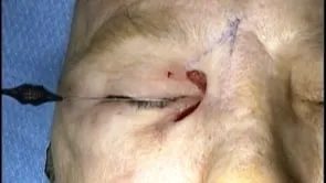
This is Richard Allen at the University of Iowa. This video demonstrates reconstruction of a relatively large defect with a median forehead flap. In addition, the defect involves the medial portion of the upper and lower lid. A periosteal strip will be used to rebuild the posterior lamella of the medial upper and lower lid and to provide support.
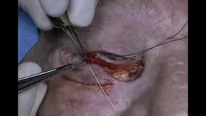
A 15 blade is used to make an incision through the periosteum and a Freer periosteal elevator is used to elevate the flap and reflect it medially. This flap will stretch to the medial edge of the upper and lower eyelid. The flap will be split centrally with Westcott scissors and then each limb of the periosteal strip will engage the upper and lower eyelid. The periosteal strip is sutured to the eyelids with 5-0 Vicryl suture. This is performed in a cross swords fashion so that the upper limb engages the lower eyelid and the lower limb engages the upper eyelid.
The forehead flap is then raised. A 15 blade is used to make an incision through the skin, subcutaneous fat and frontalis muscle to the subgaleal plane, which is superficial to the periosteum. The monopolar cautery is used to dissect in this preperiosteal plane. Approximately two centimeters superior to the orbital rims, the periosteum is then incised with a 15 blade and further dissection is carried out inferiorly in the subperiosteal plane. This plane is used to avoid damage to the neurovascular structures in this area by staying deep to them.
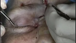
Wide undermining is then performed with the monopolar cautery at the edges of the donor site. The donor site is then closed. In doing this, the forehead flap should be transposed into appropriate position. Closure of the donor site is performed with deep interrupted 4-0 Vicryl sutures. This can sometimes be challenging due to the tension at the closure and further undermining may be necessary.
The flap is then reflected into position and sutured in position with deep interrupted 5-0 Vicryl sutures followed by a combination of 5-0 and 6-0 Prolene sutures superficially, depending on the thickness of the skin. The donor site is closed superficially with interrupted 5-0 Prolene sutures placed in a vertical mattress fashion in order to evert the skin edges. At the conclusion of the case, the flap should appear healthy. Minimal pressure should be applied to the graft postoperatively to ensure adequate perfusion.